物质科学公共实验平台X射线实验室在Bruker D8 Advance多晶X射线衍射仪上配置了高通量的90位自动进样器、全自动的DBO防散射系统及LynxEye XE-T能量分辨探测器;另外在原位XRD上,配置了高功率的转靶光源、大面积的二维面探测器Eiger及多功能的原位池;最后,高分辨粉末XRD齐备Cu/Ag靶与二维面探Eiger、兼顾毛细管透射与平板反射,也极大限度地支持高分辨粉末及PDF数据的采集与分析。上述设备及相关附件均已到货并安装,可高效、全方位支持西湖大学科研人员的相关研究工作。
X射线衍射技术是研究材料结构的重要实验工具,其结果能为功能材料的设计和改性提供重要的理论依据,而样品制备的质量直接影响XRD谱图效果,如由于不当制样可引起的实验数据误差,给结果分析带来巨大挑战。因此,了解XRD样品的观测要求、制备过程,提高样品制备质量,可以为XRD实验起到事半功倍的效果。
1.XRD样品制备要求
X射线衍射分析的样品主要有粉末样品、块状样品、薄膜样品、纤维样品等。样品不同、分析目的不同(定性分析或定量分析),则样品制备方法不同。
1.1粉末样品的制备
样品粒度和衍射强度的精确度有直接关系。粒度小,在样品架上的颗粒就越多,有足够多的颗粒被X光照射到,越接近颗粒随机分布,衍射强度就越精确;但也不能太小到纳米,使得衍射峰宽化。因此测试时要求粉末样品粒度均匀,理想的颗粒尺寸约为10-50μm。
对于颗粒较大的样品及压片烧结的材料,测试前需要通过手工或机械研磨制备合适颗粒尺寸的粉末样品;如果颗粒过粗,参与衍射的晶粒数目不够,则不能满足获得正确的粉末衍射图谱数据的条件,即样品受光照体积中晶粒的取向应该是完全随机的;如果粉末颗粒过细(<10μm),将产生对X射线的微吸收,也会降低衍射强度;当粉末颗粒太细(<100 nm)则会造成衍射峰的宽化;对于纳米材料则需将其分散均匀。
1.1.1常规粉末样品制备
实验室中常用的X射线衍射仪一般采用Bragg-Brentano衍射几何,即使用平板样品进行测试,因此要求样品的测试面平整致密,并且测试面与样品台的表面高度一致。
平板测试面,通常会采用正压法和背压法进行制样(图1和图2)。
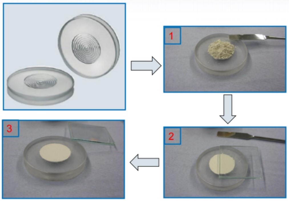
图1 正压法制样流程
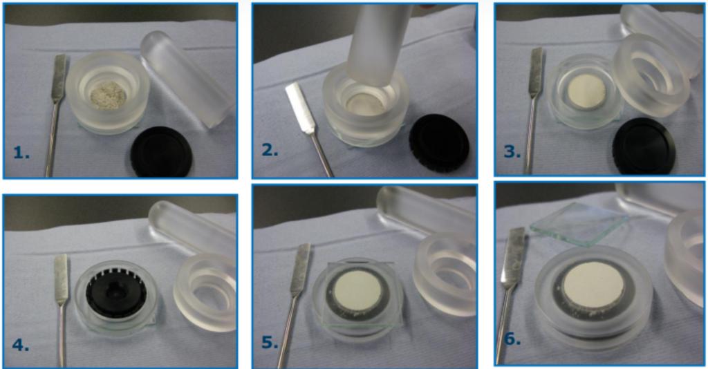
图2 背压法制样流程
1.1.2微量样品制备
制备微量样品须选用微量样品架,且将微量粉末样品分散在样品架中心(图3);极微量样品可用不溶的易挥发溶剂分散,如乙醇或丙酮等,使粉末成为薄层浆液状,均匀地涂布开来,形成一个单颗粒层的厚度就可以,待乙醇或丙酮挥发至干即可进行测量。
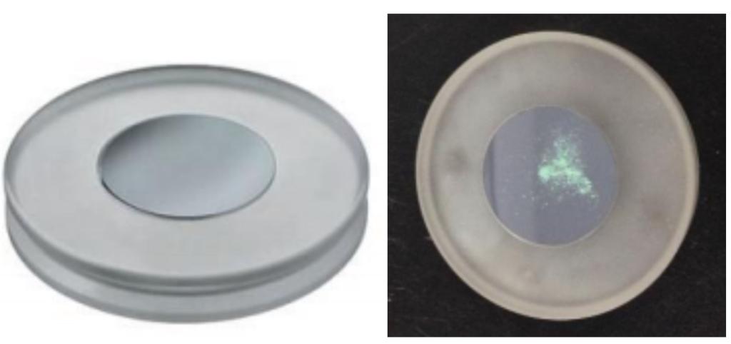
图3 微量样品架及其制样
1.2块状样品的制备
1.2.1金属块状样品
若需要了解金属自身的相组成、结构参数时,应该尽可能地磨成平面并进行简单抛光,这样不但可以去除金属表面的氧化膜,也可以就消除表面应变层;而后用超声波清洗去除表面的杂质,但样品面积尽量大于10mm×10mm。
1.2.2基片上的薄膜样品
薄膜的厚度应大于20nm,并在做测量前先确定基片的取向。将基片切割成约10mm×10mm方片后,用橡皮泥将基片垫高至与深槽样品架表面齐平(图4)。
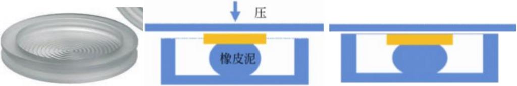
图4 深槽样品架及其制样
1.2.3不规则块体样品
对于断口、裂纹的样品,要求断口尽可能的平整并提供断口所含的元素。对于断口样品也需采用井深的深槽样品架(见图4)。
1.3纯膜/纤维样品的制备
直接将独立支撑的薄膜样品放置在零背景样品架上(见图3),保薄膜不卷曲。卷曲的薄膜需剪切成平正的小片薄片后放置在零背景样品架上。
1.4择优取向的样品制备
对于强择优取向的棒状、片状样品,可用毛细管法制样(见图5),研细的样品可随着毛细管的转动,有更多取向的小晶粒被曝光而使取向随机化,因此可以大大降低择优取向。
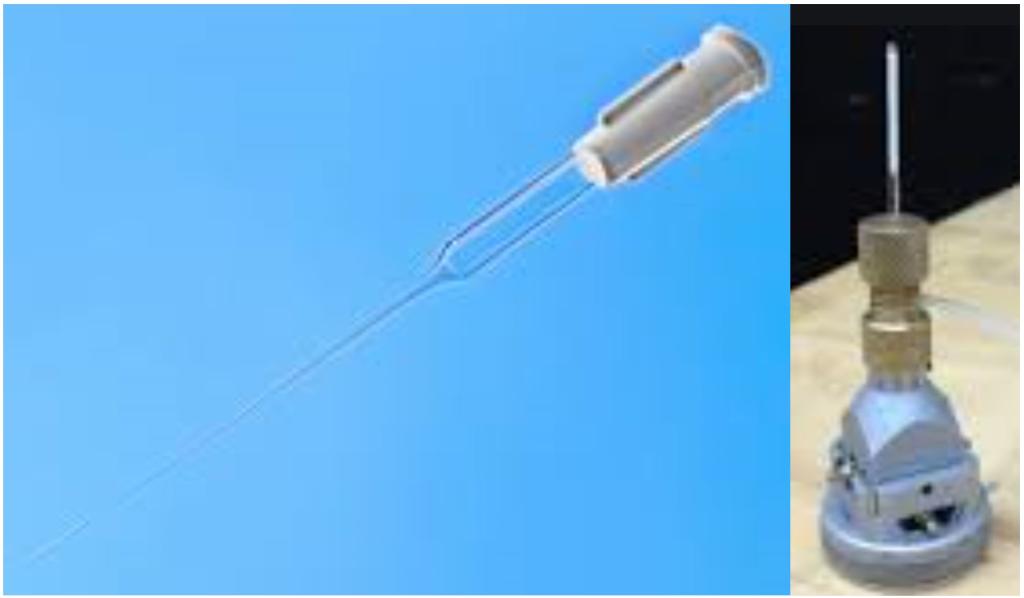
图5 毛细管法制样
2.XRD样品制备的影响因素
2.1样品表面平整度
在Bragg-Brentano几何中,因为衍射主要发生在平板样品的表面,因此样品表面的平整程度对衍射数据具有很大影响。如图6所示表面抹平样品b及未抹平的样品a的测试数据。从谱图中可以看到,样品表面不平整会导致衍射峰强度剧烈降低;同时,背底强度也 会随之下降。这一现象主要受到两个方面的影响,其一是不平整表面的漫反射会降低产生衍射的晶面数量,从而降低衍射强度;其二是在极度不平整的表面,产生的衍射峰会被相邻颗粒阻挡从而降低衍射强度。因此在制备平板样品的时候要特别注意其表面平整度。

图6 样品表面平整度对衍射测试结果的影响
2.1样品高度影响
在测试过程中,样品台的高度、样品在样品槽中的填充情况都会影响到最终样品平面相对于衍射平面的高度变化。在正常的测试过程中,样品平面应该与测试圆、衍射圆均相切,样品高度变化后,会引起测试平面与测试圆偏离相切关系,从而导致测 得的衍射峰位出现偏移。
如果测试面高于或低于样品台表面,将会引起衍射峰位的偏移与峰形的不对称,峰位偏移与高度偏差存在:Δ2θ = -(114.59*s*cosθ)/R
s =样品置位偏差(mm)R =测角仪半径(mm)
因此样品测试面须与聚焦圆相切(图7)。

图7 样品在衍射仪中的摆放位置
如图8所示,是样品台高度明显低于正常高度的情况下得到的衍射数据与正常测试情况的对比。可以发现,衍射平面的降低会造成衍射峰位的明显偏移,同时峰型也会出现一定的变化。
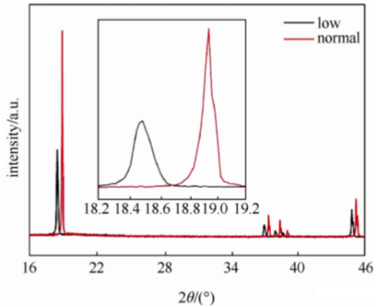
图8 样品高度对衍射测试结果的影响
2.3样品表面粗糙度
如果样品测试面比较粗糙,将会增加样品对X射线的漫散射,引起衍射峰加宽、背底增强。
2.4择优取向
样品的择优取向主要受到样品形貌的影响,通常棒状及片状的样品更容易产生择优取向,而球形样品则不易产生择优。如图9所示,是同一LiCoO2样品因制样方式不同产生的不同图谱。图9(a)是普通的正压法制备的样品,图9(b)是采用毛细管制备的样品。从图中能够看到,不同制样方式产生的衍射图谱其(003)与(104)峰的比值出现了较大的差异。
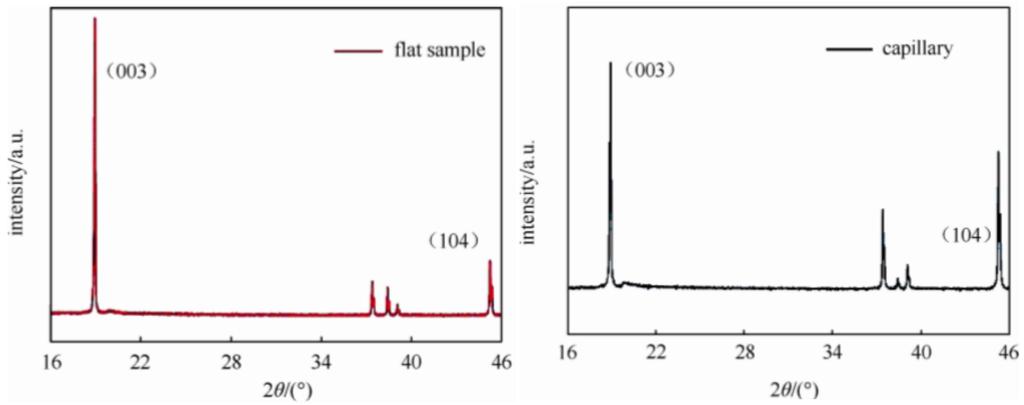
图9 (a)是普通的正压法制备的样品(b)是采用毛细管制备的样品
对于不同吸收性质的粉末,颗粒度可以认为“足够细”的尺寸要求是各不相同的,因为样品受到X射线照射的有效体积和可以忽视样品中微吸收效应的颗粒上限都取决于样品的吸收性质。Brindley对此作过详细的分析,他在衍射分析中对粉末的颗粒度按μD值进行分级(μ为物质的线吸收系数,D为晶体的平均直径)。
细 颗 粒:μD < 0.01
中等颗粒:0.01 < μD < 0.1
粗 颗 粒:0.1 < μD < 1
在Brindley的分级中,“细”表示大多数颗粒周围的吸收性质是均匀的,其差异可以忽略(微吸收效应可以忽略);对中等以上的颗粒,则需要考虑“微吸收效应”;而“十分粗”的样品,衍射实际上只局限在表面一层的晶粒;此时,粉末照片开始出现不连续的点状线。
对于X射线吸收系数很低的样品(有机物、药物等),其平均等效衍射高度低于样品的实际表面高度,造成峰位的偏差和峰型的不对称。
Δ2θ = (sin2θ)/(2µR), 其中μ是样品的线性吸收系数;R:测角仪半径(mm)
简单说来,粉末材料的高度、粗糙度、平整度、微吸收均影响着衍射峰位及强度,因此样品制备对于后续XRD实验测量起着举足轻重的作用。
Sample preparation for Powder XRD
XRD lab from ISCPS has equipped a multi-functional Powder XRD, Bruker D8 Advance, with a high-throughput 90-seat autosampler, automatic DBO optics and energy-resolved LynxEye XE-T detector;and a powerful in-situ XRD, Bruker D8 Discover, with high-brilliance rotational anode and large-area 2D Eiger detector;and also a high-resolution PXRD and PDF scattering system, with Cu and Ag anodes, large area 2D Eiger detector as well as optional stages of BB reflection or capillary stage, in-depth and full-scale to support‘corresponding scientific research at Westlake University. All the mentioned equipment have be installed in XRD Lab to facilitate high-resolution data acquisition and smooth interpretation.
XRD is an essential tool to explore the material structure, and providing solid proof to the design and property investigation of functional materials. Whereas, the sample preparation plays a key role in influencing the quality of XRD spectra. Furthermore, proper sample preparation avoids systematic errors and challenging in data interpretation. Thus, understanding and improving the sample preparation can yield double the result with half the effort.
1-Requirements for sample preparation
There are various sample forms, for instance, powder, block, thin film, fiber, liquid etc. Diverse sample preparation serves for various applications.
1.1 Powder sample preparation
Grain size is coherent with the accuracy of diffraction intensity. When it is fine grain, the more particles involved, the more chance to be irradiated, and the more closed to the random distribution, it will result in accurate diffraction intensity. However, never ground samples to nano particles, which introduces the risk of broadening peaks. Thus an ideally homogeneous grain size is 10-50μm.
Regarding large particles or calcinated materials, generally require for harsh ground, either manually or by machine, to achieve a proper grain size for diffraction measurement. Supposed that coarse particles, less suitable crystallites participating in diffraction, inaccurate diffraction intensity will be received. Namely, the orientation of crystallines in the radiating volume, should be as random as possible. But it will bring micro absorption and weaken the diffraction intensity when the particle size is smaller than 10μm; furthermore, the fine powder, with the size below 100 nm, will result in peak broadening. Hence, nano materials are better to be homo-dispersed to improve the broadening effect.
1.1.1Preparation of regular powder samples
In conventional XRD lab, Bragg-Brentano geometry (BB reflection) is most frequently applied. It desires for flat surface, which levels out at sample holder plane. There are two manners to prepare samples for BB geometry, direct assembly and back assembly, and their procedures are presented as below.
Fig 1. The procedure of direct assembly
Fig 2. The procedure of back assembly
Zero-background holders are recommended for small-amount samples, which are well-distributed at the center of the holder. Trace samples are preferred to be dispersed in bad volatile solvent, such as ethanol or acetone, to form turbid liquid and cascaded on the holder, drying out and ready for measurement. The procedure is detailed as below:
Fig 3. Zero background holder and its sample preparation
1.2 Preparation of block samples
1.2.1 Metal blocks
To understand the composition and structure of metals, flattening and polishing the sample surface is necessary, which not only removes the superficial oxidants, also eliminates the stressed top layer. Then please tidy the sample with sonication with the area above 10mm×10mm.
1.2.2 Films on substrates
The thickness of film is preferred over 20 nm, with orientation of substrate pre-confirmed. The substrate is trimmed to 10mm×10mm, supported with clay and levels out at holder surface, detailed as below.
Fig 4. Deep-well holder and its sample preparation
1.2.3 Irregular shaped samples
Samples with fracture and cracks, better to flatten it, and elemental composition is required. Deep-well holders are helpful in this case (refer to Fig 4).
1.3 Free-standing films and fiber-like samples
Directly loading the free-standing films and fiber-like samples on the zero-background holders (refer to Fig 3), ensuring a flat spread-out. Truncate a curly film into tiny pieces and sprawl on the zero-background holders.
1.4 Preparation of orientation-preferred samplesUnder this circumstance, a capillary sealing is recommended (detailed in Fig 5). As the rotation of sealed capillary, the well-ground fine powder sample can be fully exposed to X-ray, and the orientation is more close to random distribution to eliminate the preferred orientation.
Fig 5. Sample preparation in capillary
2. Impacting factors of sample preparation on diffraction
2.1 The flatness of sample surface
In BB geometry, the diffraction majorly allocates at the sample surface, and the flatness of sample surface plays a vital role in diffraction intensity. It is observed that the sample with flat (Fig 6b) and rough (Fig 6a) surface behaves dramatically differently in diffraction. It is implied that the rough surface not only weakens diffraction intensity, but also weakens the signal/noise ratio. There are two reasons are responsible for this phenomenon: firstly, rough surface generates scattering, which definitely reduces diffraction intensity; secondly, the diffracted X-ray will be blocked by adjacent particle due to coarse surface, which also lead to weak diffraction.
Fig 6. the influence of sample surface flatness in diffraction
2.2 Sample height
The holder height and the way of sample filling the holder also affect the displacement of diffraction plane. During routine measurement, the sample surface is well aligned to the measurement circle and diffraction circle. But as the sample height alters, the deviation of sample surface from the measurement circle will be introduced, and also brings the shift of diffraction peaks. Suppose that the sample surface is not equal to holder plane, it will draw into peak shift and peak shape distortion, which is well described by the formula below:
Δ2θ = -(114.59*s*cosθ)/R
in which, s: sample physical shift (mm), R: geometry radius (mm)
Fig 7. the displacement of sample loading in diffractometer
XRD scan shown in Fig 8 displays a comparison of a proper-prepared sample and mistreated sample, and the latter presents peak shift and transformation.
Fig 8. the effect of sample height in diffraction
1.1Sample surface roughness
The more rough sample surface is, the more introduction of X-ray scatter is, which also contributes to peak broadening and background enhancing.
1.2Preferred orientation
The preferred orientation is coming from sample morphology. In general, bar-like or pallet samples is prone to bear preferred orientation while spherical one is not. From Fig 9, displays (a) regular direct assembly of samples; (b) samples sealed in capillary, it’s clearly to observe the (003) and (104) peaks behave remarkably different with various sample preparation.
Fig 9. (a) regular direct assembly of samples; (b) samples sealed in capillary
As far as diverse powder absorbance is involved, the particle size varies in the requirement of fine ones, due to the effective volume exposed to the X-ray as well as the maximum micro-absorption of samples, though neglectable, depends on its total absorption property. Brindley specifically analyzed the coherence of grain size and absorption, according toμD (μ: absorption coefficient, D: the diameter of grain).
fine grains: μD < 0.01
medium grains: 0.01 < μD < 0.1
coarse grains: 0.1 < μD < 1
Among the classification ranks in Brindley’s study, fine grains can be understood as homogeneous absorption from surrounding particles and the discrepancy can be ignored (namely the micro absorption is neglectable in this case). But that above medium, we should consider the micro absorption while the effect of coarse ones should be seriously counted in, because the diffraction occurs only focusing on the superficial surface and turns out to be inconsecutive Debye ring composed of reflections.
In some low absorption samples (such as organics, medicine), their effective height is a bit lower than the actual visualized sample surface, which brings peak shift and distortion. There equation below well defines their relationship:
Δ2θ = (sin2θ)/(2µR),
In which,μ: absorption coefficient, R: the radius of grain
All in all, the factors of height, roughness, flatness and micro absorption place great emphasis on the diffraction peak and their intensity. Thus, properly preparing samples is essential in XRD data acquisition and interpretation.